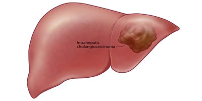
Hemangiomas are the most common tumours found in the liver. Nowadays many times they are found incidentally on ultrasound abdomen while being investigated for some other reasons. They are usually harmless and do not require any treatment; medical or surgical. Rarely they may become symptomatic and surgically treated with resection. Most of the times if you can prove convincingly that the tumour is a hemangioma you can safely forget it. A triple phase contrast CT scan or triple phase contrast MRI can confirm the diagnosis. Needle aspiration (FNAC) is usually not required.
These tumours ocuur primarily in females and associated with regular intake of Oral Contraceptive Pills (OCP). These tumours can turn into cancer in some patients. It is sometimes difficult to diagnose adenoma on the basis of imaging ( Ultasound/CT/MRI) alone. The treatment is surgical removal.
This is another type of tumour which can be diagnosed confidently with CT or MRI and can be left alone and observed. This type of tumour do not turn into cancers. Rarely in symptomatic cases surgical resection is indicated.
Hepato cellular carcinoma is a liver cancer which may develop in a healthy liver but more commonly in a cirrhotic liver. Patient developing cirrhosis due to hepatitis B/hepatitisC have more chance of having liver cancer and should have regular checkup with his doctor.
Liver cancer normally doesnot cause any sym0ptom when they are small only in advanced stage it can cause jaundice, weight loss, loss of appetite, low grade fever, lump palpable in the abdomen.
Ultrasonography, Triphasic CT scan abdomen are the test to reach the diagnosis. Some time contrast MR and bone scan are required.
Alpha fetoprotien(AFP) is tumour marker.If the AFP level in the blood is raised, it very strongly suggests that the patient has a HCC. But the converse does not always apply – if the AFP levels are normal, that does not rule out a HCC.
Needle biopsy is not mandatory but can be done in few selected cases though it carries a small risk.
It depends upon the the size of the tumour, location , number, it has spread to nodes, lungs /bones, whether liver is healthy or cirrhotic and fitness of the patient to undego operation if required.
If the liver is healthy, the patient fit, and the tumour can be safely cut out, then surgical resection of the tumour can be considered. If the tumour is large and cannot be safely removed, then chemoembolisation can be considered. If the tumour is small but the patient is unfit/unwilling to withstand surgery, radiofrequency ablation (RFA) can be considered. This involves placing a needle into the tumour and destroying it with energy generated at the tip of the needle.
If the liver is diseased (i.e. cirrhotic) but still in overall good condition, the patient is fit, and the tumour small, then liver transplantation should be considered. Surgical resection of the tumour or RFA may also be possible options. If the tumour is large and these options are not feasible, then chemoembolisation may be considered.
If the patient is unfit for surgery, the liver is very badly diseased, or the tumour is very large or has spread beyond the liver, then control of symptoms should be the main focus of care. Chemotherapy (in the form of intravenous injections or tablets taken by mouth) is not particularly effective in HCC, nor is radiotherapy.
Liver is a common site of secondary cancer i.e. Primary tumour is some where else but it has spread to liver. t is now well-recognised that surgical removal of the liver metastases from colon and rectum cancer can achieve a cure in a significant proportion of patients. When it comes to other cancers, surgical resection of liver metastases may still have a role to play, but only in a very small proportion of patients.
Another group of patients who may similarly benefit from liver resection are patients with liver metastases from neuro-endocrine tumours.
Diagnosis is usually made on the basis of an ultrasound scan, and then a CT or an MR is done to confirm that. A whole body scan called FDG PET is useful in determining if there are other secondaries elsewhere in the body.
Treatment of the liver secondaries depend upon the size of the tumour,condition of the liver, number and site of the secondaries and overall fitness of the patient.
Surgical resection of the tumour if indicated and can be done gives the best result. Other option are RFA, Chemotherapy.
In a healthy liver upto two-thirds of the liver can be removed . Liver is an organ which can regenerates itself in six to eight weeks to its original size.Only if the liver is not healthy then we tend not to remove
a large portion of the liver.
All operation has got a risk. In major liver resection the risk of mortality /death is around 5% in other way chances of survival is 95%.
To book your appointment with a gastro surgeon, please reach out to us on +91 8160650099

